


A Classification System for Loss of Papillary Height

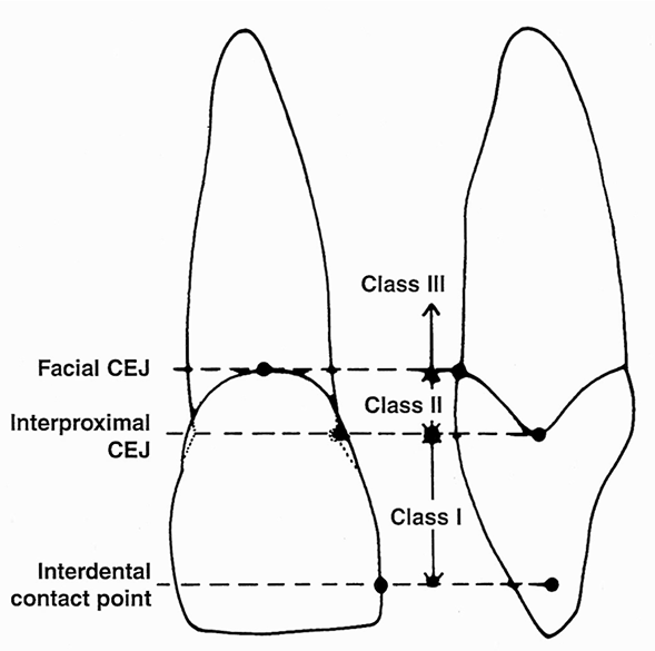
Figure : Illustration of the proposed classification system. Adapted from W. Peter Nordland,*and Dennis P. Tarnow, 1998
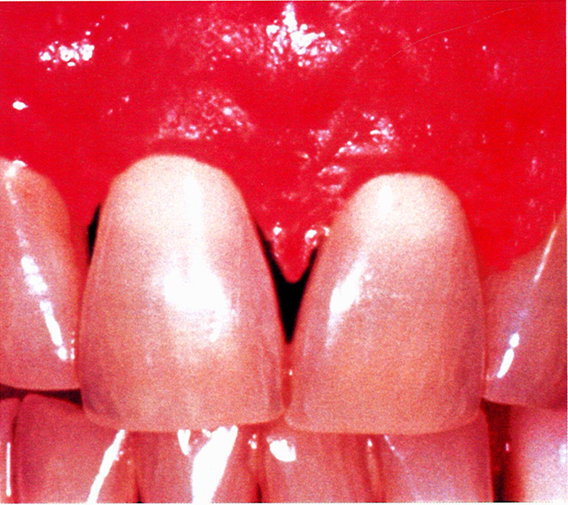
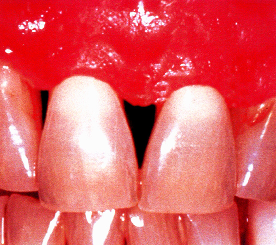
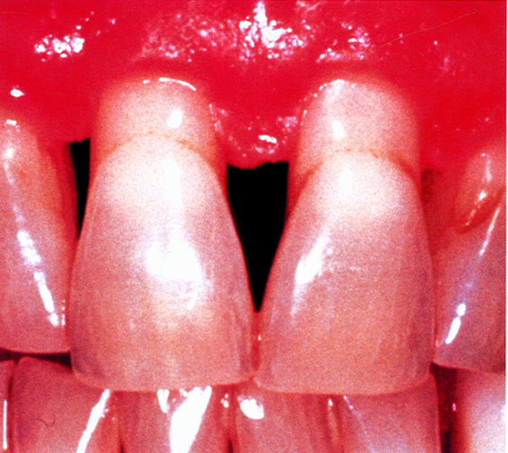
The reduction in interdental papillary height often occurs as a consequence of periodontal disease, and may also result from the healing response following periodontal therapy and the re-establishment of periodontal health.
A simple and descriptive classification system is proposed utilizing three distinct anatomical landmarks:
Interdental contact point,
Facial apical extent of the cementoenamel junction (CEJ), and
Interproximal coronal extent of the CEJ.
Normal – the interdental papilla completely fills the embrasure space up to the apical extent of the interdental contact point or area

Class I. The tip of the interdental papilla lies between the interdental contact point and the most coronal extent of the interproximal CEJ (space is present, but the interproximal CEJ is not visible).

Class II: The tip of the interdental papilla lies at or apical to the interproximal CEJ but remains coronal to the apical extent of the facial CEJ (interproximal CEJ is visible).

Class III: The tip of the interdental papilla lies at the level of or apical to the facial CEJ.
Other factors which may potentially affect the outcome of papillary augmentation procedures include the amount of interdental bone loss (Tarnow, D. P., Magner, A. W., Fletcher, P., 1992) and the width of the interdental space.
Bone levels can be recorded radiographically as the distance from the CEJ to the interdental bone crest (mean of two proximal measurements). The width of the interdental space can be measured radiographically at the level of the CEJ.